Procedures

Abdominal Aorto-iliac Occlusive Disease: Bifurcated Graft Bypass

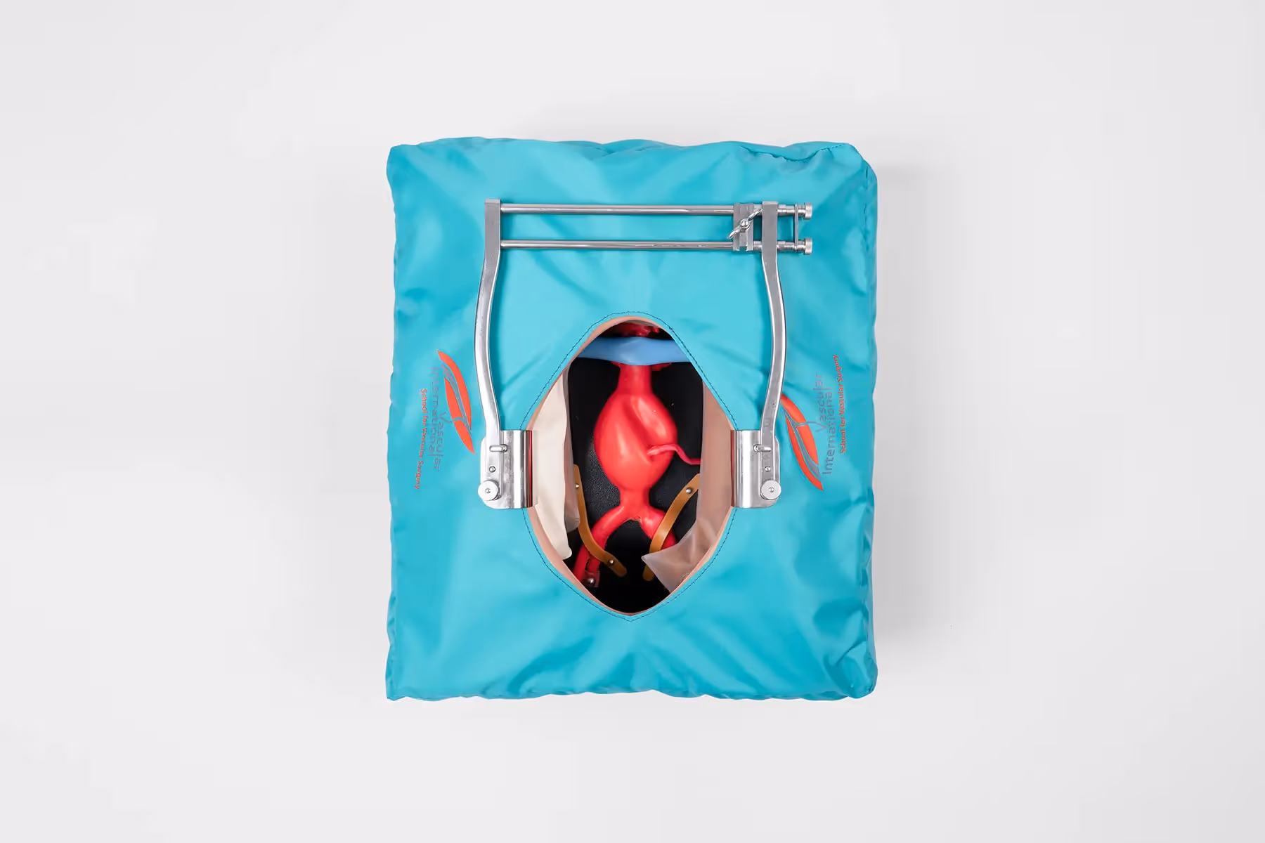
Occlusive disease of distal infrarenal aorta and/or common iliac arteries. Collateral network providing blood flow to lower limbs (lumbar arteries or inferior epigastric arteries). As treatment a aorto-bi-iliac (external iliac artery) or aorto-bi-femoral bifurcation graft bypass needs to be done to restore blood flow to the lower limbs.

Educational objectives

  • Implantation of a para-/infrarenal aortic bifurcated graft (Dacron 16x8mm or 18x9mm)
  • End-to-End (optional End-to-Side) anastomosis with clamping of one renal artery
  • To always apply intravenous heparin before arterial clamping (except patient has a history of heparin induced thrombopenia (HIT)
  • To apply tangential(partial) aortic clamp at the infrarenal aorta correctly (pay attention to orientation of calcified plaques)
  • to decide if suture line needs to be reinforced
  • To stitch correctly through arterial wall inside out (perpendicular direction)
  • To move the graft towards the correct position before pulling the thread after every stitch
  • To decide if distal anastomosis should be end-to-end or end-to-side on common iliac artery or external iliac artery
  • To rinse graft limbs and target vessels before restoring blood flow
All Steps in Detail

Step 1: Exposition

  1. Abdominal wall retractor and transperitoneal exposition of the infrarenal aorta, including definition of anatomical landmarks
  2. Placement of silicone tube around the proximal aortic neck distally to the renal arteries
  3. Preparation of distal bypass zones. Placement of vessel loops around both external iliac arteries or common femoral arteries
  4. Supra- /infrarenal crossclamping using Glover clamp (rear branch of the clamp is conducted behind the aortic wall by means of a silicone tube)
  5. Left lateral longitudinal incision (more than 3 cm long), beginning below the left renal artery
  6. Limited thrombendarterectomy of the infrarenal aorta

Step 2: Proximal anastomosis

  1. Standing on the right side of the body the suture starts at 5 o‘clock on the contralateral side (suture material: 3-0 double needle polypropylene suture)
  2. First stitch inside-out on the aorta, put a suture clamp on the second needle
  3. Second stitch with the same needle outside-in on the graft and inside-out on the aorta clockwise to first stitch
  4. Continue with a running suture around the heel up to 8 o‘clock, put a suture clamp on that needle when needle is outside the graft and pull on both clamps (suture ends) approximating graft and aorta (moving the graft up and down)
  5. After positioning the graft at the anastomotic level, continue the suture with the second needle completing the cranial wall suture of the aorta
  6. Flushing and control of the proximal anastomosis under pulsatile pressure, additional stitches if necessary (U stich, Borst stitch, suture reinforcement)

Step 3: Distal anastomosis

  1. Clamping of target vessel
  2. Longitudinal incision of 1 cm. Rounding ofincision ends with a 3.5 or 4 mm punch (allows better view into the artery)
  3. Anastomosis with bypass graft starting at 5o’clock outside-in graft and inside-out artery using 5-0 double needlepolypropylene suture.
  4. Continue suturing around the heel in parachutetechnique until 8 o’clock. Approximate the graft to the artery (mowing down thegraft)
  5. Continue suturing around the toe with otherneedle, flush and knot under pressure
  6. Perform same procedure on other side

Control of educational objectives

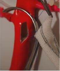
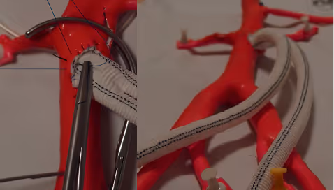
Proximal Anastomosis

Pictures and Drawings

No items found.

Courses Teaching this Procedure

European Vascular Master Class

Jan 21, 2026
 - 
Jan 23, 2026
More Information

Simulators Used for this Procedure

Abdominal Aorta Simulator

More Information

About Vascular International

The Vascular International (VI) School for Vascular Surgery is dedicated to advancing the training and education of vascular surgical techniques through the use of lifelike models. With patient safety as the top priority, VI continuously strives to enhance vascular surgery training, ensuring the highest standards of safe and effective open and endovascular patient care.

About Us